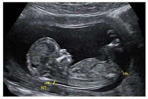
Sonogram Of Down Syndrome Fetus
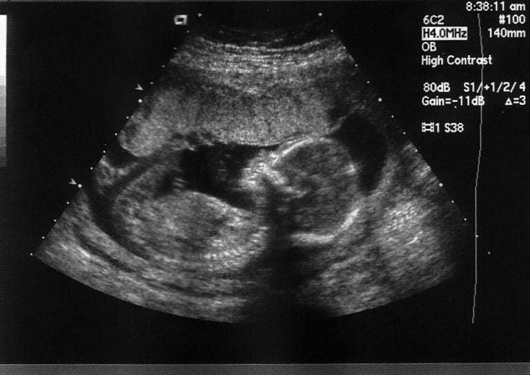
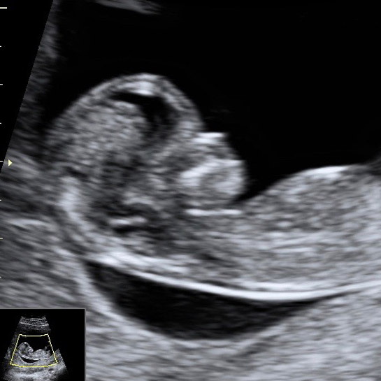
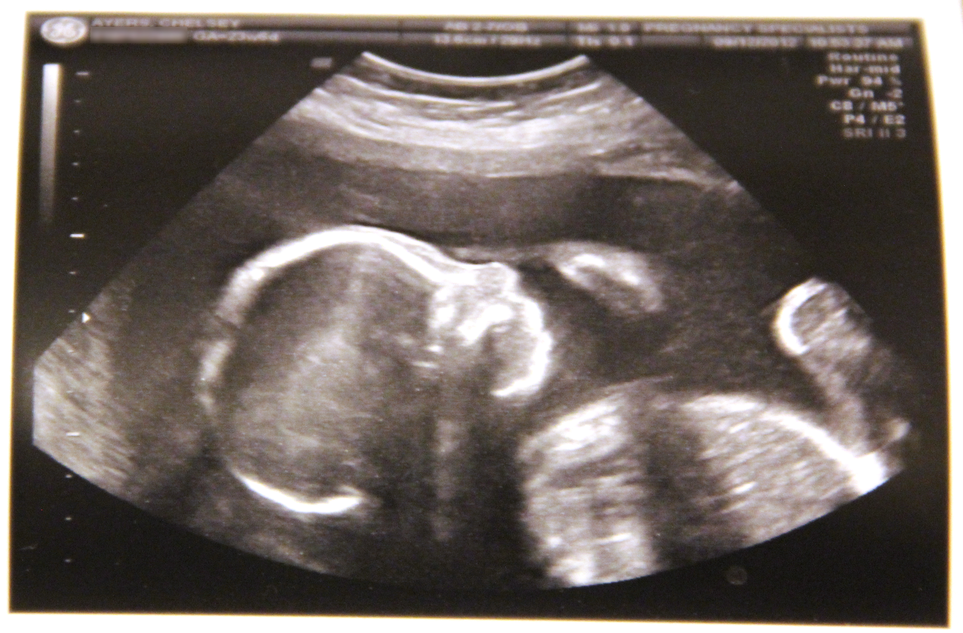
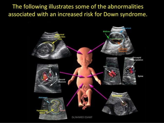
Sonogram of down syndrome fetus. In the prenatal period increased nuchal translucency hygroma colli hydrops fetus congenital heart disease kidney defects larger amount of amniotic fluid can be observed in affected fetuses with this syndrome. Down syndrome detection continues to be one of the main challenges. The FP line was defined as the line that passes through the mid-point of the anterior border of.
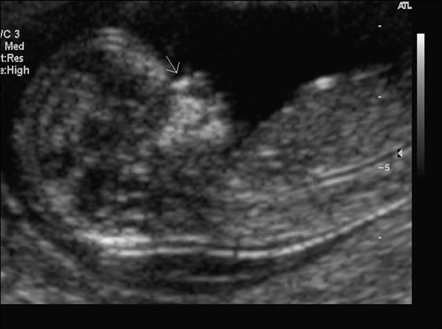
Noonan syndrome NS one of the most common RASopathies has an estimated incidence of 1 in 1000-2500 live births. There are no nasal bones and prefrontal edema is present. Both major structural abnormalities and minor soft markers can be detected by ultrasound in fetuses affected with aneuploidies.
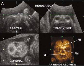
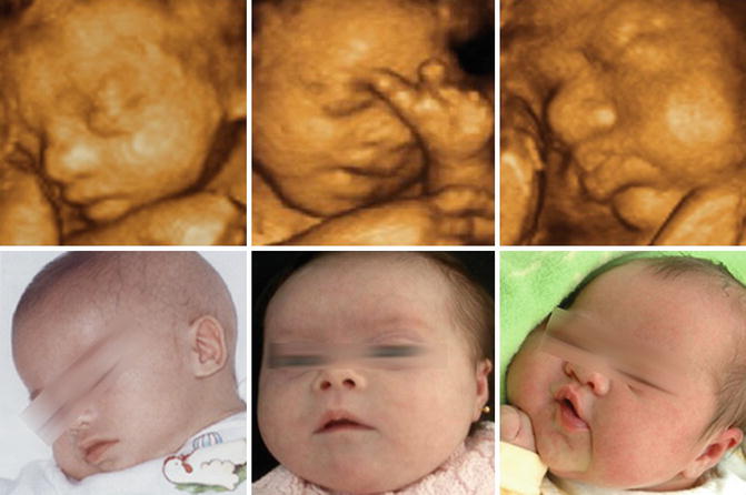
The detection can be performed by identifying various parameters from the ultrasound images recorded during the 1st 11-14 weeks and 2nd trimester 15-22 weeks of the gestational period. The nasal philtrum is very prominent due to excessive development of the upper maxilla. Two-dimensional ultrasound images in a euploid fetus at 24 6 weeks gestation a and a fetus with Down syndrome at 28 2 weeks b showing maxillanasionmandible angle.
Ultrasound can detect fluid at the back of a fetus neck which can be an indicator of down syndrome. It is done during the first trimester between 11 and 14 weeks and combines information from the ultrasound examination of the fetus and the blood of the mother. An ultrasound test measures nuchal translucency.
Interest in the vascular system in the fetus with Down syndrome was raised when it was discovered that Doppler. The combined screening test is also known as the first trimester screening test. The Genetic Sonogram is an ultrasound examination done on second trimester fetuses that not only evaluates the fetus for structural malformations but also searches for the sonographic markers of fetal Down syndrome.
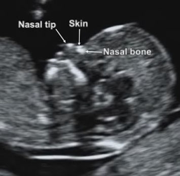
In what was called the genetic sonogram by researchers such as B. Thickened nuchal fold nuchal translucency Duodenal Atresia double bubble Echogenic bowel Cardiac heart anomalies Choroid plexus cyst Echogenic intracardiac focus. Currently Down syndrome is identified visually by looking at the ultrasonogram image of the fetus for the presence of nasal bone.
Down Syndrome can include cardiovascular central nervous craniofacial musculoskeletal gastrointestinal and urinary tract system anomalies. Early detection of Down syndrome is important and significant for a better assessment of the fetus.
However they are seen more frequently in fetuses with an abnormality.
This is an effective method in the early detection of health disorders. The fetus presents at 26 weeks micromelia in one arm with monodactyly and facial dysmorphism. The FP line was defined as the line that passes through the mid-point of the anterior border of. The answer to that question is yes. Currently Down syndrome is identified visually by looking at the ultrasonogram image of the fetus for the presence of nasal bone. Approximately 30 of babies with Down syndrome have detectable abnormalities on the mid-trimester ultrasound 1. The combined screening test is also known as the first trimester screening test. Soft markers are sonographic findings that do not in themselves cause any adverse outcomes. The nasal philtrum is very prominent due to excessive development of the upper maxilla.
One soft marker that might have shown up on the first-trimester NT screening which is always performed between weeks 10 and 13 is nuchal-fold thickening where the area at the back of a babys neck accumulates fluid causing it to appear thicker than usual. Currently Down syndrome is identified visually by looking at the ultrasonogram image of the fetus for the presence of nasal bone. Both major structural abnormalities and minor soft markers can be detected by ultrasound in fetuses affected with aneuploidies. Interest in the vascular system in the fetus with Down syndrome was raised when it was discovered that Doppler. The Genetic Sonogram is an ultrasound examination done on second trimester fetuses that not only evaluates the fetus for structural malformations but also searches for the sonographic markers of fetal Down syndrome. This is an effective method in the early detection of health disorders. It is done during the first trimester between 11 and 14 weeks and combines information from the ultrasound examination of the fetus and the blood of the mother.






/babyboyultrasound-7bf2ced4b4794754b67dea974b7ec744.jpg)








Post a Comment for "Sonogram Of Down Syndrome Fetus"