Popliteal Artery Entrapment Syndrome Radiology
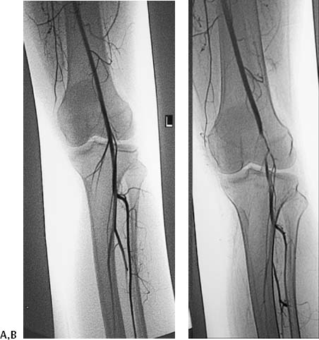
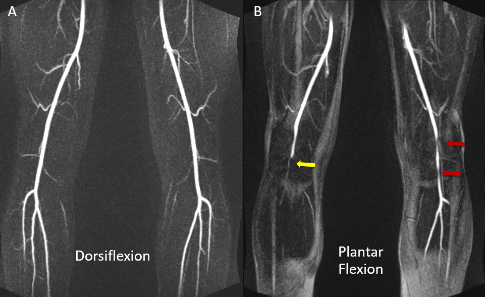
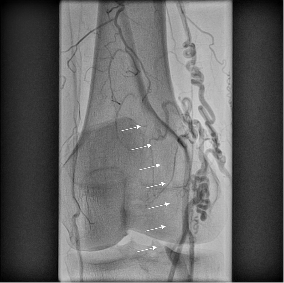
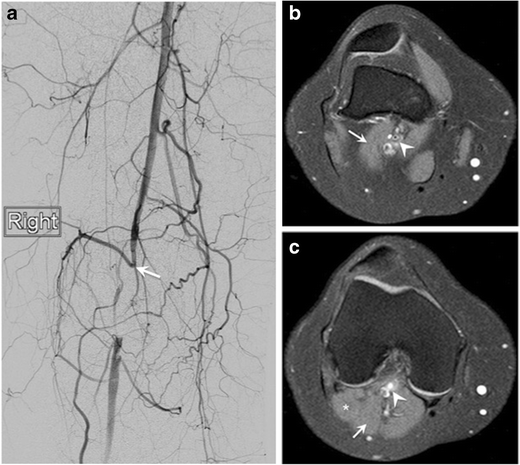
Popliteal artery entrapment syndrome radiology. Popliteal artery entrapment syndrome. B Repeat selected arteriogram with the foot in active plantar flexion shows extrinsic compression causing complete occlusion. 19341895 Indexed for MEDLINE Publication Types.
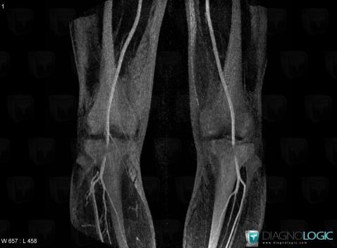
Popliteal artery entrapment syndrome is a rare abnormality of the anatomical relationship between the popliteal artery and adjacent muscles or fibrous bands in the popliteal fossa. Case Study - Presentation. Popliteal artery entrapment syndrome is an uncommon entity typically affecting young athletic males who present with symptoms of calf claudication.
Popliteal artery entrapment syndrome is due to either an acquired or a congenital abnormality 20. Popliteal artery entrapment syndrome. Sectional radiological and threedimensional images are useful for not only depiction of the arterial changes but also identification of the abnormal anatomic structures responsible for the entrapment.
2 Computed Tomographic Angiography and Digital Subtraction Angiography Findings in Popliteal Artery Entrapment Syndrome. 820 Jorie Blvd Suite 200. However multislice CT provides detailed information on the wall diameter of the artery and relation of the artery to adjacent structures by a non-invasive technique.
Despite multiple consultations the patient was not diagnosed with this syndrome for over 10 years leading to occlusion of his left popliteal artery. Surg Radiol Anat 1995. Rosset E Hartung O Brunet C et al.
Popliteal artery entrapment syndrome. 17161 169 Google Scholar. Housseini AM1 Maged IM Abdel-Gawad EA Hagspiel KD.
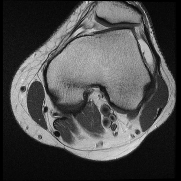
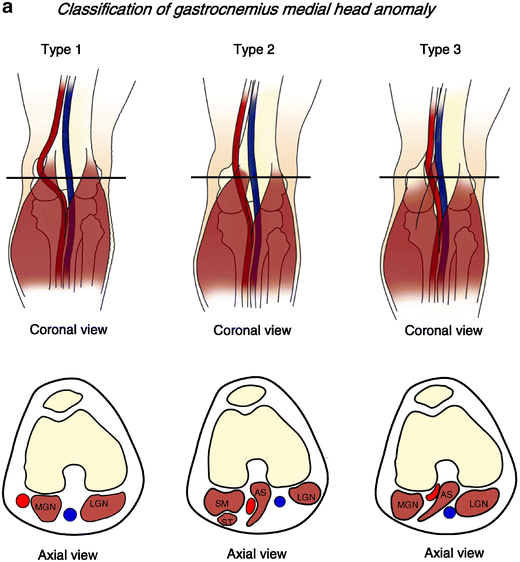
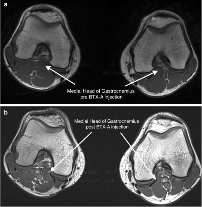
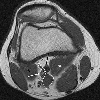
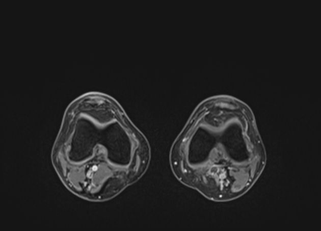
The diagnosis of popliteal artery entrapment syndrome may be confirmed by Doppler ankle pressures3 20 pulse volume recordings duplex Doppler26 35 36 37 computerized axial scanning38 magnetic resonance imaging23 25 39 40 or magnetic resonance angiography. A predisposing anatomical anomaly is seen with the popliteal artery located medial to the medial head of gastrocnemius MHG and separate to the popliteal vein.
Despite multiple consultations the patient was not diagnosed with this syndrome for over 10 years leading to occlusion of his left popliteal artery.
Popliteal artery entrapment syndrome. 19341895 Indexed for MEDLINE Publication Types. Housseini AM1 Maged IM Abdel-Gawad EA Hagspiel KD. In radiology the combination of radiological examination not only develops the function and anatomical state of the popliteal artery but also the detailed structures of popliteal fossa can be visualized. 8ara H Sugano T Fuji N. Popliteal artery entrapment syndrome is an uncommon entity typically affecting young athletic males who present with symptoms of calf claudication. Surg Radiol Anat 1995. B Repeat selected arteriogram with the foot in active plantar flexion shows extrinsic compression causing complete occlusion. 820 Jorie Blvd Suite 200.
Popliteal artery entrapment syndrome. A predisposing anatomical anomaly is seen with the popliteal artery located medial to the medial head of gastrocnemius MHG and separate to the popliteal vein. Housseini AM1 Maged IM Abdel-Gawad EA Hagspiel KD. Popliteal artery entrapment syndrome. Evaluation with spiral CT. Popliteal artery entrapment syndrome. It is caused by an anomalous relationship of muscle and artery in the popliteal fossa resulting in extrinsic arterial compression.















Post a Comment for "Popliteal Artery Entrapment Syndrome Radiology"